Showing posts with label Pediatrics. Show all posts

CTEV (Congenital Talipes Equinovarus) or Clubfoot

Overview
clubfoot is a general term used to describe a common deformity in which the feet change / bend of the state or the normal position. Some of foot deformities including talipes deformity of the ankle called the talus derived from the word (which means ankle) and plague (which means the foot). Deformities of the foot and ankle are sorted depending on the position of the ankle and foot abnormalities. Talipes deformity are:

  • Talipes varus: inversion or bend into the>
  • Talipes valgus: eversion or bending outward
  • Talipes equinus: plantar flexion of the fingers which is lower than the heel
  • Talipes calcaneus: dorsiflexion where the radius is higher than a heel
clubfootMost clubfeet which is a combination of several positions and the highest incidence rate is the type of talipes equinovarus (TEV) in which the foot down and into the arched position with varying degrees of severity. Unilateral clubfoot is more common than other types of bilateral and can occur as abnormalities associated with other syndromes such as chromosomal aberration, artrogriposis (general immobility of the joints), cerebral palsy or spina bifida.

Frequency of clubfoot from the general population is 1: 700 to 1: 1000 live births in which the boy two times more often than women. Based on the data, 35% occurred in monozygotic twins and only 3% in dizygotic twins. This emphasizes the role of genetic factors

Pathophysiology
The exact cause of clubfoot is not known until now. Some experts say that the disorder was caused by an abnormal position or limited movement in the womb. Another expert said that the abnormality occurs due to abnormal embryonic development of the current developments towards flexion and foot eversion at month 7 of pregnancy. Growth is impaired in these phases will cause a deformity which is influenced also by the intrauterine pressure.

Evaluation of diagnostic
This deformity can be detected early during the prenatal ultrasonography or detected at birth.

Therapeutic management
Rapid growth during the period of the infant allows for the handling of remodeling. Treatment begins when abnormalities are obtained and consisted of three stages, namely:
1) correction of deformity,
2) correction to maintain normal muscle balance is achieved,
3) observation and follow-up to prevent the return of the deformity.
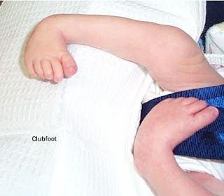
clubfood, CTEV, CTEV Foot, CTEV Treatment, clubfoot club, the clubfoot, clubfoot treatment, clubfoot deformity, what is clubfoot,clubfoot surgery, congenital clubfoot
Correction of CTEV is with manipulation and serial application of the "cast" that starts from birth and continue until the goal is achieved correction. This correction is supported also by stretching exercises of the structure of the medial side of foot and exercise a weak contraction of the structure on the lateral side. Manipulation and the use of "cast" is repeated on a regular basis (from several days to 1-2 months at intervals of 1-2 months) to accommodate the rapid growth in this period. If manipulation is not effective, surgical correction to improve the structure of the redundant, extend or tendon transplant. Then ektremitas will be in the "cast" until the goal is achieved correction.

Prognosis
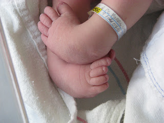
clubfood, CTEV, CTEV Foot, CTEV Treatment, clubfoot club, the clubfoot, clubfoot treatment, clubfoot deformity, what is clubfoot,clubfoot surgery, congenital clubfoot
Some cases showed a positive response to treatment, whereas some other cases show that the response time or do not respond at all to Treatment. Parents should be informed that the results of the Treatment is not always predictable and depends on the severity of the deformity, the child's age at intervention, the development of bones, muscles and nerves. Long-term foot function after Treatment is generally good but the study results show that the correction as adults will show a 10% foot smaller than usual.

Tags : CTEV Foot, CTEV Treatment, clubfoot club, the clubfoot, clubfoot treatment, clubfoot deformity, what is clubfoot,clubfoot surgery, congenital clubfoot

Anemia In Infants


Anemia is a disease characterized by too few red blood cells (erythrocytes) in the blood.

Causes
Anemia in the newborn may result from:
  • Blood loss
  • Destruction of red blood cells are excessively
  • Impaired red blood cell formation.

The loss of large amounts of blood during the birth process can occur if the placenta is detached from the uterine wall prematurely (placental abruption) or if there is a tear in the umbilical cord.
Infants looked very pale, low blood pressure and shortness of breath.

Anemia in premature infants is usually caused by blood loss (due to repeated blood tests for laboratory tests) and a reduced red blood cell formation.
Under normal circumstances, the bone marrow did not form new red blood cells for 3-4 weeks after birth. Anemia will worsen as the baby's growth rate is faster than the rate of formation of new red blood cells. But premature babies usually do not show symptoms of anemia and this situation will disappear by itself within 1-2 months.

Destruction of red blood cells occurs in:
  • Hemolytic disease in newborns: a large number of red blood cells are destroyed by antibodies produced by the mother during the fetus in the womb
  • Babies with deformities of the red blood cells, such as spherocytosis (spherical red blood cells)
  • Abnormalities of hemoglobin (oxygen carrying protein in red blood cells), such as sickle cell disease or thalassemia
  • Infection for the baby in the womb (eg toxoplasmosis, German measles, cytomegalovirus disease, herpes simplex or syphilis).
If blood cells are destroyed, hemoglobin is converted to bilirubin. High levels of bilirubin in the blood (hyperbilirubinemia) causes jaundice and in severe cases, can cause brain damage (kern icterus).

Anemia due to iron deficiency can occur in infants aged 3-6 months if given cow's milk or infant formula is not fortified with iron.

Treatment
  • If blood loss occurs during the birth process, immediately given a blood transfusion.
  • If the cause is the destruction of red blood cells is excessive, replace transfusion, where the baby's blood is replaced with fresh blood. Red blood cells are damaged, bilirubin and antibodies from the mother's body dumped.
  • In iron deficiency anemia are given extra iron.
  • If symptoms of severe anemia, a blood transfusion.
Tags : Anemia infants, anemia in babies, anemia in children, anemia babies treatment

    Biliary Atresia


    Biliary atresia is a condition where the bile ducts are not formed or does not develop normally. Biliary atresia is a liver disease in children who contributed to 50-60% of liver transplants. The function of the biliary system is to remove metabolic wastes from the liver and transports bile salts needed to digest fats in the small intestine. In biliary atresia there clogging the flow of bile from the liver to the gallbladder. This can cause liver damage and cirrhosis of the liver, which if untreated can be fatal. The incidence of biliary atresia throughout the world 1: 10,000 live births. Biliary atresia is a liver disease in children who contributed to 50-60% of liver transplants.

    Causes
    The cause is a combination of various things such as viral infections especially reovirus and rotavirus, genetic disorders, toxic material that interferes with the growth of the biliary tract and the presence of perinatal bile duct damage during delivery.

    Symptoms
    Symptoms usually occur within 2 weeks after birth, which are:
    • Dark-colored urine baby
    • Pale stools
    • Yellow skin
    • Do not gain weight or slow weight gain
    • Enlarged liver.

    By the time the baby reaches the age of 2-3 months, will arise the following symptoms:
    • Impaired growth
    • Itching
    • Fussy
    • High blood pressure in the portal vein (blood vessels that carry blood from the stomach, intestines and spleen to the liver).

    Type of biliary atresia
    There are 3 (three) types of biliary atresia:
    1. Type I, atresia of the common bile duct;
    2. Type II atresia of the duct while hepatikus
    3. Type III obstruction or blockage of the bile duct more upstream in the liver tissue at the porta hepatis and the channel above the porta hepatis.
    Most patients with biliary atresia, including the type III, which is as much as 90%.

    Pathophysiology

    Pathophysiology of biliary atresia is not known with certainty. According to figures histopatologik, it is known that biliary atresia occurs because of prolonged inflammatory processes that cause extrahepatic biliary ducts are progressively damaged. In the advanced state of the inflammatory process spread to the intrahepatic biliary ducts, so it will experience a progressive damage.

    Diagnosis
    Diagnosis based on symptoms and physical examination.
    On abdominal examination, palpable enlarged liver.

    Regular checks carried out:
    • Blood tests (there are elevated levels of bilirubin)
    • abdominal ultrasound
    • X-ray abdomen (liver appears enlarged)
    • Cholangiography
    • Liver biopsy
    • laparotomy (usually done before the baby is 2 months old).

    Treatment

    Kasai operation, according to the name of the Japanese doctor who discovered the technique of the operation, then the technique was performed bypass surgery which makes bile duct from the liver directly into the duodenum or the duodenum. Successful Kasai operation, particularly in type I biliary atresia and a little on the type II. Whereas in type III, the Kasai operation is not very satisfactory results. As a result, the disease will progress to cirrhosis of the liver in which liver texture is soft and supple previously become hard like stone.
    In all organic disease that has reached the final stage, there is no other way to treatment than do organ transplants in this case the liver or liver transplant. Special liver transplant in infants is actually relatively easy to find a donor, which is one of the parents, could be the father or mother of the baby. Depending on the results of examination of whether the father or mother of the most close and match blood groups and other test results such as MHC type I, type II MHC, etc..


    Tags : biliary atresia procedure, kasai, biliary atresia kasai, kasai procedure, extrahepatic biliary atresia, congenital biliary atresia, biliary atresia symptoms, neonatal biliary atresia, biliary cirrhosis, emedicine biliary atresia, biliary atresia symptoms,