Fever - Type, Cause, Sign and Symptom, and Treatment

Image from kobieta.pl
What is a fever?
Fever is the body temperature rising abnormally. Febrile or fever is generally interpreted in body temperature above 37.2 º C.

Range of different types of fever
Type of fever that may be encountered include:
1. Septic fever
Temperature gradually rose to the level that high once at night and fall back to the level above normal in the morning. Complaints are often accompanied by chills and sweating. When high fever is down to the level of a normal fever is also called hectic.
2. Fever remittances
Body temperature can go down every day but never reached normal body temperature. Causes may be recorded temperatures can reach two degrees and no temperature differences were noted for septic fever.
3. Intermittent fever
Body temperature dropped to the level of normal for several hours in a day. When the fever as it occurs within two days once called tersiana and if there are two free days of fever between the two bouts of fever called kuartana.
4. Continuous fever
Temperature variations throughout the day did not differ by more than one degree. At the level of persistent high fever once called hyperpyrexia.
5. Cyclic fever
An increase in body temperature for several days, followed by a period free of fever for several days followed by a rise in temperature as before.
A type of fever is sometimes associated with a particular disease such as type of intermittent fever for malaria. A patient with symptoms of fever may be connected immediately with an obvious cause such as abscesses, pneumonia, urinary tract infections, malaria, but sometimes simply can not be connected immediately with an obvious cause. In practice 90% of the patients with fever who had just experienced, is primarily a self-limiting illness such as influenza or other similar viral diseases. But this does not mean we do not have to remain vigilant against bacterial infection.

Causes of fever
The cause of fever besides infection can also be caused by circumstances toxemia, malignancy or a reaction to the drug, also in disorders of the central temperature regulation center (eg, cerebral hemorrhage, coma). Basically to achieve the required accuracy of diagnosis of the causes of fever include: accuracy of patient medical history taking, physical examination execution, observation of the disease course and laboratory evaluation. and other appropriate support and holistic.
Some specific things to look for in a fever is a fever ways, the old fever, high fever as well as complaints and symptoms that accompany fever lian.
Undiagnosed fever is a condition where a patient has a fever continuously for 3 weeks and the body temperature above 38.3 degrees Celsius and still has not been obtained even though the cause has been studied intensively for one week by means of laboratory and other medical support.

Sign and Symptoms
1. Body temperature over 37.2º C
2. Sweating
3. Elevated Respiratory
4. Shivering


Therapy
1. Antipyretic
2. Antibiotics, according to program
3. Avoid alcohol or ice pack

The Process of Menstruation


Menstruation is the periodic and cyclic bleeding from the uterus caused by the collapse of the endometrial tissue. The process of menstruation takes place with the following steps:

a. Menstrual stage or desquamasi

At the stage of menstruation, from the womb through the vagina with menstrual bleeding and the endometrial lining of the cervical mucus. The blood that comes out does not freeze because there fermen that prevents blood clotting and dilute mucosal pieces.
If your period is a lot of the fermen insufficient causing menstrual blood clots. The amount of bleeding during menstruation is normally ± 50 cc. At the stadium menstrual endometrium becomes thin.


b. Post Menstruum Stadium or Regeneration Stadium

This stage since the day of the fourth period is called the endometrium due to injuries that are released gradually re-covered by a mucous membrane of the epithelium of the newly-endometrial glandular epithelium ± 0.5 mm.

c.  Inter Prolifer Stadium or  Menstruum Stadium

This stage gradually from day 5 of menstruation until day 14 of the first day of menstruation. Glands grow more rapidly than other tissues at this stage of ± 3.5 mm thick endometrium.

d. Pre Menstrual Stadium  or  Secretion Stadium

At this stage endometrial thickness remained unchanged but the form of the gland into a long and winding as well as removing sap. Secretory stage lasts from days to 14-28.

Warm-Hot compresses


Definition of Compress

Compres: a pad of cloth applied firmly to a part of the body; compres may be dry or wet, cold or warm (Smith, 1996).

Based on the above definition that the pack can be supplied in dry or wet and cold or warm. Using a hot compress media can be a bag of hot water / hot water bottle, hot steam, mud, hot towels, electric pads and others.

Benefits / effects of heat

Heat is used extensively in the treatment because it has great effects and benefits. The benefits / effects of heat are (Gabriel, 1996):
a. Physical Effects
Heat can manyebabkan liquid, solid and gas experience expansion in all directions.
b. Effects of Chemical
Heat can increase the speed of chemical reactions. Metabolism in tissues will occur with increasing exchanges between the body of chemicals with body fluids.
c. Biological Effects
Heat can cause dilation of blood vessels that lead to increased blood circulation.
In fisilogis the body's response to heat causes dilation of blood vessels, lowering blood viscosity, reduce muscle tension, increase metabolism and increase the permeability of the capillary network. The response of the heat is used for therapeutic purposes in various conditions and circumstances that occur in the body.

Mechanism of Heat

Heat loss or energy enters the body through the skin in four ways: conduction, convection, radiation and evaporation (Gabriel, 1996).
The working principle of a warm compress by using a hot jar wrapped in a cloth that is the conduction heat transfer which occurs from the bladder into the body so that it will cause the dilation of blood vessels, so it will be a decrease in muscle tension. Compress is performed using hot jar wrapped in a cloth, temperature 36° -38° C, which is placed directly on the right side and left side alternately abdomen between the stomach and right hand side of the left abdomen every 5 minutes for 20 minutes. Hot water is replaced every five to ten minutes to maintain the temperature of the hot jar to keep warm. Compress given until the pain is reduced or lost (Perry and Potter, 1993).

Urinary Tract Infection (UTI)

Image from drugs.com
What is a urinary tract infection?
Urinary tract infection (UTI) is a term used to express the infasi microorganisms in the urinary tract.
Glossary:
1. Significant Asymptomatic bacteriuria (ASB) is a significant bacteriuria without symptoms.
2. Bacterial Cystitis is a syndrome consisting of:
a. Urinating less time.
b. Frequent urination (day and night).
3. Abacterial Cystitis (urethra Syndrome) is a syndrome consisting of:
a. Urinating less time.
b. Frequent urination without any bacteria in the bladder.

What causes urinary tract infection?
The most common cause of UTI is a Gram-negative bacteria that normally inhabit including intestine which then rises into the urinary system. Of gram negative turns E. Coli occupies the top spot, followed by Proteus, Klebsiela, Enterobacter, and Pseudomonas.
This type of Gram-positive Coccus less frequently as a cause of UTI, while Enterococcus and Stapilococcus aureus is often found in patients with urinary tract stones, elderly men with prostate hipertrophi or in patients using catheters. If found S. aureus in the urine should be suspected hematogenous infection of the kidney. So it is with pseudomonas aeroginosa can infect the urinary tract of hematogan path and in approximately 25% of patients with isolated tipoid salmonilla in the urine. Bacteria that can cause a UTI by way haematogenous is brusela, nokardia, aktinormises, and mycobacterium tuberkolosae.
Viruses are also found in urintanpa symptoms of acute UTI. Adenovirua types 11 and 12 suspected as cause of hemorrhagic cystitis. Hemorrhagic cystitis may also be caused by Scistosoma hematobium flatworms that belong to this class. Candida is a fungus that most commonly cause UTIs, especially in patients with catheters, diabetes mellitus or patients who received treatment with antibiotics spktrum area.


What are the symptoms of urinary tract infections?
Clinical symptoms of UTI are not typical and even in some patients without symptoms. Symptoms are often found is dysuria, polaki Syria, and urinary urgency that usually occur together. Supra pubic pain and pelvic area. Polikisuria occur due to the bladder can not accommodate more than 500 ml urine because of the inflamed mucosa, so frequent urination. Stranguria is a difficult urination accompanied by muscle spasms and waist are often found in acute cystitis. Tenesmus is pain with the desire to empty the bladder was empty though. Nocturia is likely to frequent urination at night due to decreased kandungkemih capacity. Often secondary nocturnal enuresis also found that wetting in adults, the difficulty in initiating urination prostatismus and less heavy flow of urine. Pain urethra, ureter and renal colic.
Clinical symptoms of UTI in accordance with the urine of infected as follows:
  1. At the bottom of the UTI, patients usually complain of pain or burning in the urethra during urination with little urine and discomfort in the supra pubic area.
  2. At the top of the UTI can be found symptoms of headache, malaise, nausea, vomiting, fever, chills, malaise, or low back pain.

What checks are carried out on urinary tract infections?
1. Urinalysis
a. Leukosuria
b. Hematuria
2. Bacteriological
a. Microscopic
b. Bacterial culture
3. Chemical tests
4. Plat-dye test (Dip-slide)
5. Radiological examination and other examinations.

CTEV (Congenital Talipes Equinovarus) or Clubfoot

Overview
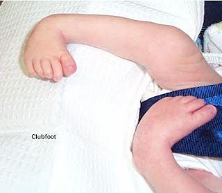
clubfoot is a general term used to describe a common deformity in which the feet change / bend of the state or the normal position. Some of foot deformities including talipes deformity of the ankle called the talus derived from the word (which means ankle) and plague (which means the foot). Deformities of the foot and ankle are sorted depending on the position of the ankle and foot abnormalities. Talipes deformity are:

  • Talipes varus: inversion or bend into the>
  • Talipes valgus: eversion or bending outward
  • Talipes equinus: plantar flexion of the fingers which is lower than the heel
  • Talipes calcaneus: dorsiflexion where the radius is higher than a heel
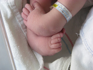
clubfootMost clubfeet which is a combination of several positions and the highest incidence rate is the type of talipes equinovarus (TEV) in which the foot down and into the arched position with varying degrees of severity. Unilateral clubfoot is more common than other types of bilateral and can occur as abnormalities associated with other syndromes such as chromosomal aberration, artrogriposis (general immobility of the joints), cerebral palsy or spina bifida.

Frequency of clubfoot from the general population is 1: 700 to 1: 1000 live births in which the boy two times more often than women. Based on the data, 35% occurred in monozygotic twins and only 3% in dizygotic twins. This emphasizes the role of genetic factors

Pathophysiology
The exact cause of clubfoot is not known until now. Some experts say that the disorder was caused by an abnormal position or limited movement in the womb. Another expert said that the abnormality occurs due to abnormal embryonic development of the current developments towards flexion and foot eversion at month 7 of pregnancy. Growth is impaired in these phases will cause a deformity which is influenced also by the intrauterine pressure.

Evaluation of diagnostic
This deformity can be detected early during the prenatal ultrasonography or detected at birth.

Therapeutic management
Rapid growth during the period of the infant allows for the handling of remodeling. Treatment begins when abnormalities are obtained and consisted of three stages, namely:
1) correction of deformity,
2) correction to maintain normal muscle balance is achieved,
3) observation and follow-up to prevent the return of the deformity.
clubfood, CTEV, CTEV Foot, CTEV Treatment, clubfoot club, the clubfoot, clubfoot treatment, clubfoot deformity, what is clubfoot,clubfoot surgery, congenital clubfoot
Correction of CTEV is with manipulation and serial application of the "cast" that starts from birth and continue until the goal is achieved correction. This correction is supported also by stretching exercises of the structure of the medial side of foot and exercise a weak contraction of the structure on the lateral side. Manipulation and the use of "cast" is repeated on a regular basis (from several days to 1-2 months at intervals of 1-2 months) to accommodate the rapid growth in this period. If manipulation is not effective, surgical correction to improve the structure of the redundant, extend or tendon transplant. Then ektremitas will be in the "cast" until the goal is achieved correction.

Prognosis
clubfood, CTEV, CTEV Foot, CTEV Treatment, clubfoot club, the clubfoot, clubfoot treatment, clubfoot deformity, what is clubfoot,clubfoot surgery, congenital clubfoot
Some cases showed a positive response to treatment, whereas some other cases show that the response time or do not respond at all to Treatment. Parents should be informed that the results of the Treatment is not always predictable and depends on the severity of the deformity, the child's age at intervention, the development of bones, muscles and nerves. Long-term foot function after Treatment is generally good but the study results show that the correction as adults will show a 10% foot smaller than usual.

Tags : CTEV Foot, CTEV Treatment, clubfoot club, the clubfoot, clubfoot treatment, clubfoot deformity, what is clubfoot,clubfoot surgery, congenital clubfoot

Anemia In Infants


Anemia is a disease characterized by too few red blood cells (erythrocytes) in the blood.

Causes
Anemia in the newborn may result from:
  • Blood loss
  • Destruction of red blood cells are excessively
  • Impaired red blood cell formation.

The loss of large amounts of blood during the birth process can occur if the placenta is detached from the uterine wall prematurely (placental abruption) or if there is a tear in the umbilical cord.
Infants looked very pale, low blood pressure and shortness of breath.

Anemia in premature infants is usually caused by blood loss (due to repeated blood tests for laboratory tests) and a reduced red blood cell formation.
Under normal circumstances, the bone marrow did not form new red blood cells for 3-4 weeks after birth. Anemia will worsen as the baby's growth rate is faster than the rate of formation of new red blood cells. But premature babies usually do not show symptoms of anemia and this situation will disappear by itself within 1-2 months.

Destruction of red blood cells occurs in:
  • Hemolytic disease in newborns: a large number of red blood cells are destroyed by antibodies produced by the mother during the fetus in the womb
  • Babies with deformities of the red blood cells, such as spherocytosis (spherical red blood cells)
  • Abnormalities of hemoglobin (oxygen carrying protein in red blood cells), such as sickle cell disease or thalassemia
  • Infection for the baby in the womb (eg toxoplasmosis, German measles, cytomegalovirus disease, herpes simplex or syphilis).
If blood cells are destroyed, hemoglobin is converted to bilirubin. High levels of bilirubin in the blood (hyperbilirubinemia) causes jaundice and in severe cases, can cause brain damage (kern icterus).

Anemia due to iron deficiency can occur in infants aged 3-6 months if given cow's milk or infant formula is not fortified with iron.

Treatment
  • If blood loss occurs during the birth process, immediately given a blood transfusion.
  • If the cause is the destruction of red blood cells is excessive, replace transfusion, where the baby's blood is replaced with fresh blood. Red blood cells are damaged, bilirubin and antibodies from the mother's body dumped.
  • In iron deficiency anemia are given extra iron.
  • If symptoms of severe anemia, a blood transfusion.
Tags : Anemia infants, anemia in babies, anemia in children, anemia babies treatment

    Biliary Atresia


    Biliary atresia is a condition where the bile ducts are not formed or does not develop normally. Biliary atresia is a liver disease in children who contributed to 50-60% of liver transplants. The function of the biliary system is to remove metabolic wastes from the liver and transports bile salts needed to digest fats in the small intestine. In biliary atresia there clogging the flow of bile from the liver to the gallbladder. This can cause liver damage and cirrhosis of the liver, which if untreated can be fatal. The incidence of biliary atresia throughout the world 1: 10,000 live births. Biliary atresia is a liver disease in children who contributed to 50-60% of liver transplants.

    Causes
    The cause is a combination of various things such as viral infections especially reovirus and rotavirus, genetic disorders, toxic material that interferes with the growth of the biliary tract and the presence of perinatal bile duct damage during delivery.

    Symptoms
    Symptoms usually occur within 2 weeks after birth, which are:
    • Dark-colored urine baby
    • Pale stools
    • Yellow skin
    • Do not gain weight or slow weight gain
    • Enlarged liver.

    By the time the baby reaches the age of 2-3 months, will arise the following symptoms:
    • Impaired growth
    • Itching
    • Fussy
    • High blood pressure in the portal vein (blood vessels that carry blood from the stomach, intestines and spleen to the liver).

    Type of biliary atresia
    There are 3 (three) types of biliary atresia:
    1. Type I, atresia of the common bile duct;
    2. Type II atresia of the duct while hepatikus
    3. Type III obstruction or blockage of the bile duct more upstream in the liver tissue at the porta hepatis and the channel above the porta hepatis.
    Most patients with biliary atresia, including the type III, which is as much as 90%.

    Pathophysiology

    Pathophysiology of biliary atresia is not known with certainty. According to figures histopatologik, it is known that biliary atresia occurs because of prolonged inflammatory processes that cause extrahepatic biliary ducts are progressively damaged. In the advanced state of the inflammatory process spread to the intrahepatic biliary ducts, so it will experience a progressive damage.

    Diagnosis
    Diagnosis based on symptoms and physical examination.
    On abdominal examination, palpable enlarged liver.

    Regular checks carried out:
    • Blood tests (there are elevated levels of bilirubin)
    • abdominal ultrasound
    • X-ray abdomen (liver appears enlarged)
    • Cholangiography
    • Liver biopsy
    • laparotomy (usually done before the baby is 2 months old).

    Treatment

    Kasai operation, according to the name of the Japanese doctor who discovered the technique of the operation, then the technique was performed bypass surgery which makes bile duct from the liver directly into the duodenum or the duodenum. Successful Kasai operation, particularly in type I biliary atresia and a little on the type II. Whereas in type III, the Kasai operation is not very satisfactory results. As a result, the disease will progress to cirrhosis of the liver in which liver texture is soft and supple previously become hard like stone.
    In all organic disease that has reached the final stage, there is no other way to treatment than do organ transplants in this case the liver or liver transplant. Special liver transplant in infants is actually relatively easy to find a donor, which is one of the parents, could be the father or mother of the baby. Depending on the results of examination of whether the father or mother of the most close and match blood groups and other test results such as MHC type I, type II MHC, etc..


    Tags : biliary atresia procedure, kasai, biliary atresia kasai, kasai procedure, extrahepatic biliary atresia, congenital biliary atresia, biliary atresia symptoms, neonatal biliary atresia, biliary cirrhosis, emedicine biliary atresia, biliary atresia symptoms,